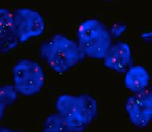
In situ hybridization (ISH) is a technique used to detect and localize the presence or absence of a specific genetic sequence in tissue using a probe with a complementary polynucleotide sequence. We offer both fluorescent (FISH) and chromogenic (CISH) in situ hybridization assays, which are available on formalin-fixed, paraffin embedded tissue slides. Dentaturing and hybridization of probes are maintained using an automated Dako Hybridizer.
Fluorescence in situ hybridization (FISH) uses fluorescent probes that bind to only those parts of the chromosome with which they show a high degree of sequence complementarity. Fluorescence microscopy can be used to find out where the fluorescent probe is bound to the chromosomes.
Chromogenic in situ hybridization (CISH) is a cytogentic technique that provides genetic information in the context of tissue morphology using immunohistochemistry techniques.
Our in situ hybridization services can meet your needs with:
- Expert sample and/or slide preparation to enhance sensitivity and signal for ISH applications
- Consultation on probe design and experiment design.
Probes will be designed using proprietary algorithms developed by experts, which allows for the best results in the most cost-effective way.


